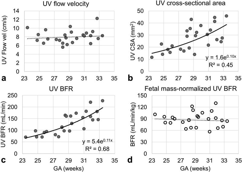
Figure 6.
Plots of the parameters used to extract fetal mass-normalized blood flow rate (BFR, mL/min/kg). The regions of interest of the umbilical vein used to estimate (A) flow velocity (cm/s) and (b) cross-sectional area (mm2) were manually traced on phase-contrast (PC) MRI images. (c) Blood flow rate (mL/min) was estimated as the product of flow velocity and cross-sectional area and (d) was normalized to fetal mass.

