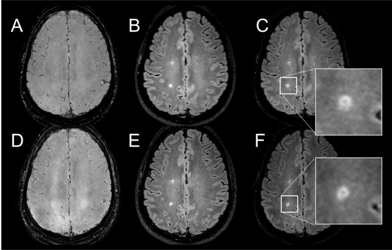
Fig. 3.

A reformatted axial slice in the brain of an MS patient showing 3D T2*-weighted gradient echo (A) and FLAIR (B) images acquired separately. The corresponding interleaved images are shown in D and E. The corresponding reconstructed FLAIR* images are shown in C and F. A vein running through a periventricular lesion is visible on both FLAIR* images (arrows), and in their magnified views (C, F).
