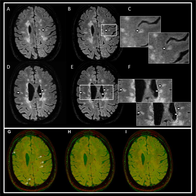
Fig. 4.

Upper panel: representative FLAIR* images from two MS patients acquired with separate FLAIR and T2* acquisitions (A, D) and the corresponding images from the interleaved sequences (B, E). The arrows point to perivenous lesions. Magnified views in (C, F) depict the lesion areas for FLAIR* (upper left) and the interleaved FLAIR* (lower right) images. The bottom panel shows an image overlay of FLAIR (red) on T2* (green) in one MS patient. The separate acquisitions (G) shows small differences in head position (arrows), which disappear with image registration (H). The images acquired with the interleaved pulse sequence are automatically aligned (I).
