
Figure 1.
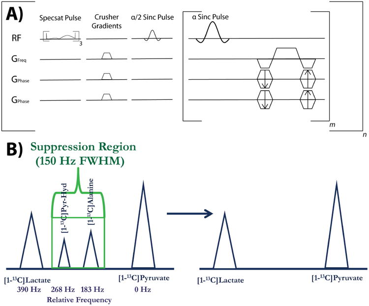
Depiction of pulse sequence and spectral suppression region. (A) Pulse sequence diagram for the 3D bSSFP sequence used in these studies, with the spectral suppression (Specsat) pulses and crusher gradients being played out 3 times and one time, respectively, prior to imaging. The sequence consists of m phase encodes and n time-points, with the 2D version sequence featuring one fewer set of phase encode gradients for projection imaging. (B) The suppression region for the spectral suppression pulse is shown here in the context of suppressing the two main peaks between [1-13C]pyruvate and [1-13C]lactate, [1-13C]alanine and [1-13C]pyruvate-hydrate. The relative frequency separation in Hz for 3T is shown below the spectrum.