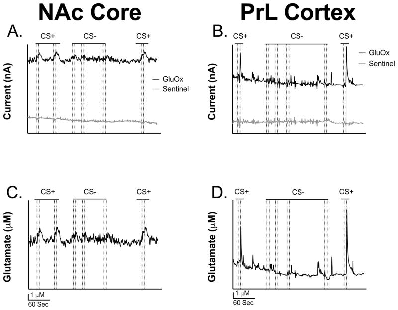
Figure 4. Representative glutamate traces from the NAc core and PrL cortex.
(A) Representative trace of glutamate release (nA) in the NAcC before sentinel site subtraction. (B) Representative trace of glutamate release (nA) in the PrL before sentinel site subtraction. (C) Representative trace of the concentration of glutamate release (μM) in the NAcC after sentinel site subtraction. (D) Representative trace of the concentration of glutamate release (μM) in the PrL after sentinel site subtraction. Note that the dotted lines represent lever extension and retraction, respectively.

