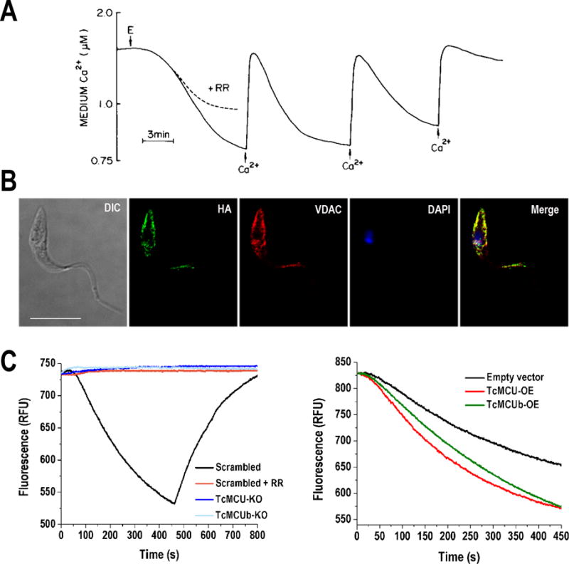
Figure 1. Mitochondrial calcium uniporter (MCU) in Trypanosoma cruzi.

A) Mitochondrial Ca2+ uptake by digitonin-permeabilized T. cruzi epimastigotes (E) in the presence of succinate. T. cruzi mitochondria has the ability to take up Ca2+ in successive additions and as in mammalian cells, mitochondrial Ca2+ uptake by MCU is inhibited by ruthenium red (RR). B) Fluorescence microscopy of endogenously tagged TcMCU-3xHA in T. cruzi epimastigotes confirms the localization of TcMCU in mitochondria. TcMCU-3xHA was detected with monoclonal anti-HA antibodies (green) whereas anti-TbVDAC (T. brucei voltage dependent anion channel) was used as mitochondrial marker (red). Yellow signal in merge indicates co-localization. Nucleus and kinetoplast were labeled with DAPI (blue). Bars, 10 μm. C) Ca2+ uptake by digitonin-permeabilized TcMCU- and TcMCUb-knockout (KO) cells. Scrambled, scrambled control cells in absence or presence of 5 μM ruthenium red (RR). The reaction was started after adding 50 μM digitonin in the presence of 5 mM succinate and 20 μM CaCl2. The variation of Calcium Green-5N fluorescence is showed in relative fluorescence units (RFU), in which a decreasing extramitochondrial Ca2+ concentration (or increasing mitochondrial [Ca2+]) is observed as a decrease in fluorescence. Addition of the uncoupler carbonyl cyanide p-trifluoromethoxylhydrazone (FCCP) releases mitochondrial Ca2+ taken up, which is evidenced by the increase of fluorescence. D) Ca2+ uptake by digitonin-permeabilized TcMCU- and TcMCUb-overexpressing (OE) and control (epimastigotes transfected with empty pTREX vector) cells. Conditions were as panel C. The image in Panel A has been reproduced with permission of (Docampo and Vercesi, 1989a).
