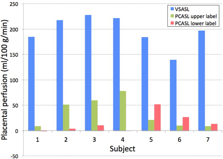
Figure 1.

Apparent placental perfusion measured using VSASL and PCASL with labeling plane above and below the imaging slab. VSASL showed dramatically higher perfusion measurement than either of PCASL methods in all subjects. With PCASL, upper labeling showed higher placental perfusion signal in the placenta that was located relatively lower in the uterus (Subject 1–4) and lower labeling showed higher placental perfusion signal in the placenta that was located relatively higher in the uterus (Subject 5–7).
