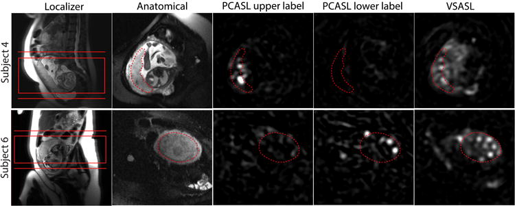
Figure 2.

Comparison of placental ASL images acquired using different labeling methods as well as localizer and anatomical images in Subject 4 and 6 from Figure 1. As shown in the localizer, the placenta was located relatively lower in Subject 4 and higher in Subject 6 inside the uterus. The common imaging slab of VSASL and PCASL and the two different locations of the labeling plane of PCASL are shown in the localizer images. In the other images, the placenta is delineated with the dotted line. PCASL signal in the placenta was higher with upper labeling in Subject 4 and with lower labeling in Subject 6, compared to the other location of the labeling plane. For both subjects, the average VSASL signal in the placenta was substantially higher than either PCASL scheme.
