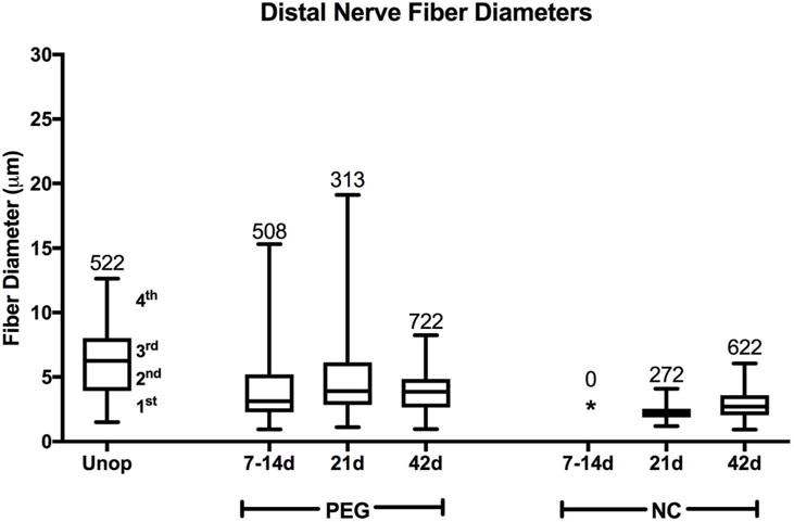
Figure 7.

Box plots showing range of nerve fiber diameters (μm) for each quartile. Plots show range of fiber diameters in Unoperated (Unop) sciatic nerves and in PEG-fused (PEG) and Negative Control (NC) sciatic nerves at 7–14d, 21d, and 42d PO for transected nerves 6mm distal to the lesion site. The line separating the 2nd and 3rd quartiles shows the median diameter. * signifies no surviving axons. Numbers at the top of the highest quartile give total number of nerve fibers counted for that protocol at that time (also see Table 2). Note that PEG-fused fiber diameters more closely resemble Unoperated than Negative Control diameters and that no Negative Control axons are present at 7–14d PO.
