Figure 9.

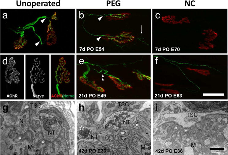
Confocal and TEM images of soleus NMJs. Unoperated (a,d), singly cut PEG-fused (b,e), and negative control (NC) (c,f) animals at 7 and 21d PO, showing SV2 and 2H3 labeled axons (green) and bungarotoxin labeled AChRs (red). Scale bar a–c, e,f = 50μm. g,h,i: TEM images of soleus NMJs from Unoperated (g), PEG-fused (h), and Negative Control (i) at 42d PO. Scale bar g–i = 2μm. Confocal images show robustly labeled nerve terminals directly apposed to their AChRs in both the Unoperated and 7 and 21d PO PEG-fused NMJs, but only a few thin axons in the 21d NC. These regenerating axons do not make contact with post-synaptic receptors. In a–f, arrowheads = single innervation, thin arrows = nerve terminal sprouts, double arrowhead = multiple innervation, and in g–i, NT = nerve terminal, TSC= terminal Schwann cell, M= muscle mitochondria, asterisks = secondary receptor folds, open star = engulfed axon material inside a Schwann cell.
