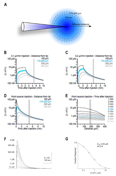
Figure 1.
Diffusion of D-APV from the injection site. A. Analytical simulations were used to evaluate the D-APV concentration at different distances from the injection site. B, C. Diffusion analysis demonstrates that, after a single D-APV injection at 0.1 μl/min (B) or 0.2 μl/min (C), there is a 10-fold dilution of D-APV at 175–200 μm from the point of injection. D. Time course of the D-APV concentration obtained when approximating the release of D-APV from the injectrode as an instantaneous event from a point source. E. Distribution of D-APV concentration within a 400 μm radius from the injection site, at different times after the injection. Slight inaccuracies in the distance between the location of the injectrode and the recording site do not confound our estimates of the effective D-APV concentration in the neuropil, measured 10 min after the injection. F. Simulations of NMDAR-EPSCs using a kinetic scheme after pre657 equilibration with different D-APV concentrations (0 nM to 10 μM) with instantaneous release of 1 mM glutamate. Glutamate transient decays exponentially with a time constant of 1 ms; G. The EC50 for blockade of NMDARs by D-APV was approximately 0.5 mM as shown in the relative response vs. concentration of D-APV curve. Because the EC50 was lower than the concentration predicted to be reached by the diffusion analysis within the stipulated time window, the chosen dose of D-APV can be assumed to effectively block >50% of the NMDARs during this time.

