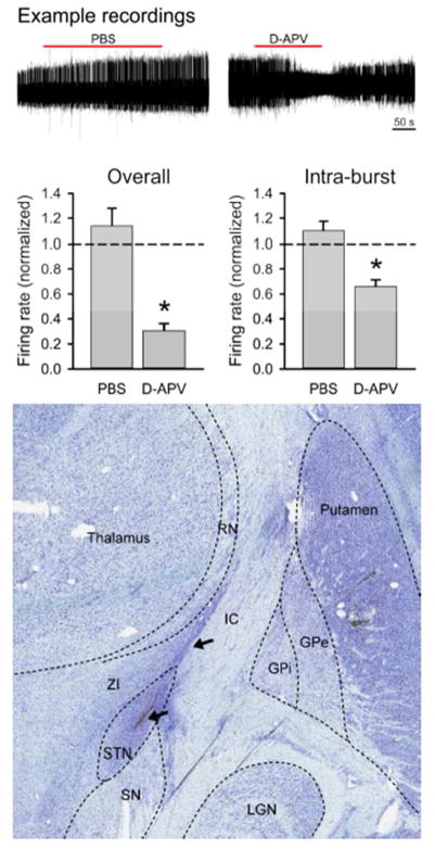
Figure 2.
Effects of D-APV on spike firing in the primate subthalamic nucleus (STN). The top row shows representative extracellular in vivo recordings of the spiking activity of STN neurons before, during and after the microinjection of 0.5 μl of saline/0.1% DMSO (left) or D-APV 5 mM/0.1% DMSO (right). The bar graph on the left shows the average responses of firing rates to injection of saline or 5 mM D-APV, expressed as a ratio of the pre-injection firing rate in individual cells. The bar graph on the right shows the average intra-burst frequencies following the injections. Data are from 11 experiments in which the vehicle was injected, and 4 cells recorded after exposure to D-APV, respectively. *, p < 0.05, t-tests, examining differences between saline vehicle and D-APV experiments. Lower row shows Nissl stained coronal section of the brain at approximately A10. The outlines of relevant surrounding brain structures are marked with dashed lines. An injection system tract is visible (arrows). Abbreviations: STN, subthalamic nucleus; SN, substantia nigra; ZI, zona incerta; GPe, external pallidal segment; GPi, internal pallidal segment; RN, reticular nucleus of the thalamus.

