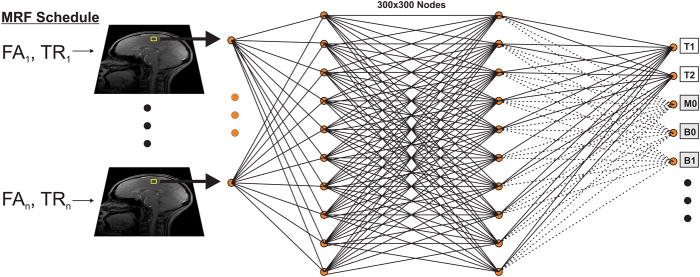
Fig. 1.
Schematic of the reconstruction approach used in this study. MRI data acquired with the optimized MRF EPI sequence is fed voxelwise to a four layer neural network containing two 300×300 hidden layers. The network is trained by a dictionary generated with the Extended Phase Graph algorithm with the tanh and sigmoid functions used as activation functions of the first and last hidden layers respectively. The network then outputs the underlying tissue parameters T1 and T2. Additional tissue parameters including M0, B0, B1 etc… (gray boxes) can similarly be obtained by training the network with a suitable dictionary.

