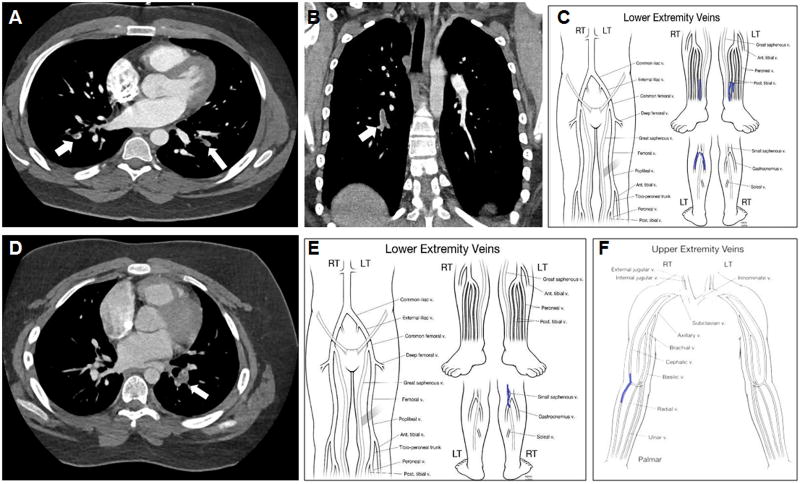
FIGURE 1.
(A) Chest CT, axial view, revealing a pulmonary embolism (arrows) in case 1. (B) Chest CT scan, coronal view, revealing an acute right pulmonary artery embolism (arrow) in case 2. (C) Lower extremity mapping computed from ultrasound revealing thromboses in bilateral posterior tibial veins and left upper calf gastrocnemius vein (case 2). (D) Pulmonary embolism on chest CT scan in case 3 (arrow). (E) Lower extremity mapping computed from ultrasound revealing a right saphenous vein thrombus in case 3. (F) Upper extremity mapping demonstrating bilateral cephalic vein thromboses in case 3.

