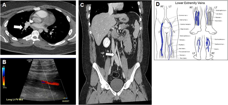
FIGURE 2.
Case 4 (A) Pulmonary embolism on chest CT (arrow); (B) Initial deep venous thrombosis on ultrasound; (C) Mild compression of the left common iliac vein as it passes under the right common iliac artery (arrow); (D) Lower extremity mapping computed from ultrasound revealing an extensive right LE deep venous thrombosis (second event).

