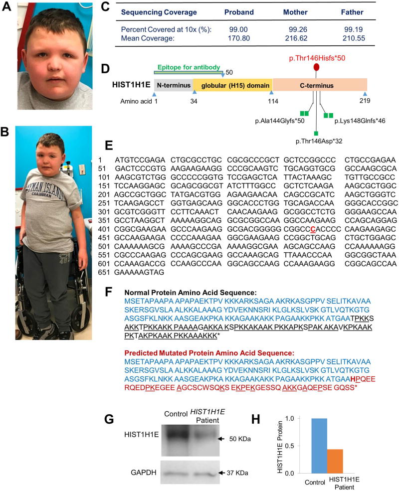
Figure 1. Dysmorphic facial and physical features of the patient.
A. Facial features. B. Full-body photograph displays relative overweight. C. Sequencing depth and coverage captured through Whole Exome Sequencing. D. Schematic illustrating the domains of the HIST1H1E protein and mutations. Locations of previously-reported mutations are illustrated with green squares, while the patient presented in this case study is represented with a red circle. The epitope used for figures G and H is against the N-terminus of the protein, and targets amino acids 1 – 50. E. Nucleotide sequence for human HIST1H1E gene. The bold, red, underlined nucleotide illustrates the patient’s c.435dupC mutation. F. The normal amino acid sequence for HIST1H1E (top) has a C-terminus (black) largely composed of K, A, and P (underlined) residues. The predicted mutated amino acid sequence (bottom) is predicted to change at the insertion point (bold), drastically reducing the content of K, A, and P residues (underlined) found in the C-terminus (red). G. Western blot using whole-cell lysate from patient and control blood samples shows a reduction in HIST1H1E protein expression. H. Quantification of western blot examining HIST1H1E protein content.

