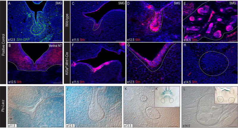
Figure 3. Shh pathway is active during early SMG development.

(A) Anti-GFP immunostaining (green) on e12.5 Shh-GFP SMG (dotted white line). (B) Anti-Shh immunostaining (red) on e12.5 ventral neural tube (dotted white line). (C-H) Anti-Shh immunostaining (red) on e11.5, e12.5, and e14.5 wild-type and Kif3af/f;Wnt1-Cre mutant SMGs (dotted white lines). Nuclei counterstained with Hoechst. (I-L) X-gal staining on frontal sections of Ptc-LacZ embryos. Dotted black lines indicate developing SMGs. (K’, L’) High-magnification images of K and L. (A, B, C, E, F, H, I) 100μm; (D, G, J) 50μm; (K, L) 500μm; (K’, L’) 200μm.
