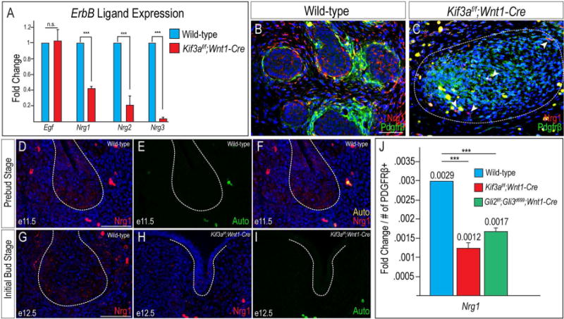
Figure 5. Nrg1 expression is reduced in ciliopathic and GliA mutants.

(A) RT-qPCR for Egf, Nrg1, Nrg2, and Nrg3 transcript levels in wild-type SMGs and Kif3af/f;Wnt1-Cre mutant mesenchymal capsules. (B, C) Anti-Nrg1 (red) and anti-Pdgfrβ (green) co-immunostaining on e14.5 wild-type SMGs and Kif3af/f;Wnt1-Cre mutant mesenchymal capsules (dotted white lines). White arrow heads in C indicate a small number of Nrg1-positive cells in the Kif3af/f;Wnt1-Cre mutant mesenchymal capsules. (D) Anti-Nrg1 immunostaining (red) on frontal section of a wild-type e11.5 SMG (dotted white lines). (E) Auto-fluorescence of red blood cells from the 488 filter in D. (F) Merged images from D and E. (G, H) Anti-Nrg1 immunostaining (red) on frontal sections of e12.5 wild-type and Kif3af/f;Wnt1-Cre SMGs (dotted white line). Nuclei counterstained with Hoechst. (I) Auto-fluorescence of red blood cells from the 488 filter in H. (J) RT-qPCR for Nrg1 transcript levels in wild-type, Kif3af/f;Wnt1-Cre, and Gli2f/f;Gli3d699;Wnt1-Cre SMGs or mutant mesenchymal capsules. Fold change of transcript levels were normalized to Pdgfrβ+ cells. ***=p<0.001 Scale bars= (B-I) 50μm.
