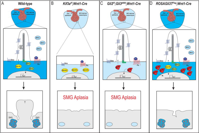
Figure 7. Hypothesized model for primary cilia and GliA-dependent SMG development.

(A) In response to Shh binding to Ptc in wild-type SMG mesenchymal cells, Gli2/3FL is shuttled through the primary cilia and processed into GliA. GliA moves to the nucleus to induce transcription of downstream genes (either directly or indirectly; dotted black line), possibly including Nrg1. SMG organogenesis occurs. (B) In Kif3af/f;Wnt1-Cre mutant embryos, cilia are lost on the NCC-derived mesenchyme and Gli2/3FL cannot be processed into GliA. Nrg1 expression is not induced and SMG aplasia occurs. (C) In Gli2f/f;Gli3f/f;Wnt1-Cre mutant embryo, primary cilia remain, but functional GliA is not produced. Nrg1 expression is not induced and SMG aplasia occurs. (D) In ROSAGli3TFlag;Wnt1-Cre mutant embryos, primary cilia remain, but an excessive amount of GliR are produced. Functional GliA is still produced and moves to the nucleus to induce transcription of downstream genes (either directly or indirectly; doted black line), possibly including Nrg1. SMG organogenesis occurs. T, tongue.
