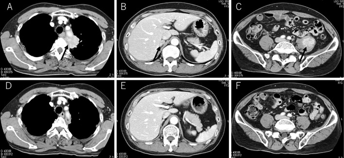
Figure 1.
CT scans showed a primary lesion in the left upper lobe (black arrow), enlarged right adrenal gland (black arrow) and enlarged left iliopsoas (black arrow) with contrast enhancement (A-C). CT images showed a reduction in the primary and metastatic lesions at the 70th dose of nivolumab (D-F).

