Figure 2.
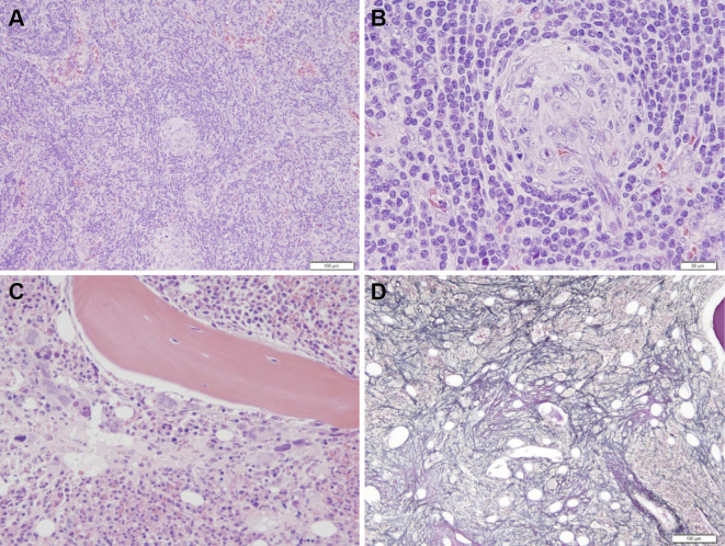
The histopathological findings. (A) and (B) The histological appearance of the left cervical lymph node. (A) Hematoxylin and Eosin (H&E) staining, 40×. (B) H&E staining, 200×. The lymph node showed interfollicular expansion and atrophic germinal centers penetrated by blood vessels. (C) and (D) The histological appearance of the bone marrow. (C) H&E staining, 200×. The examination of the bone marrow revealed hyperplastic marrow with increased megakaryocytes. (D) Silver staining, 100×. Reticulin fibrosis was observed.

