Figure 3.

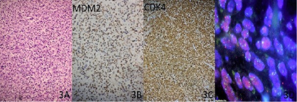
A, Light Microscopic Appearance of De-differentiated Liposarcoma; B and C, MDM2 and CDK4 Staining; D, MDM2 Amplification by FISH

A, Light Microscopic Appearance of De-differentiated Liposarcoma; B and C, MDM2 and CDK4 Staining; D, MDM2 Amplification by FISH