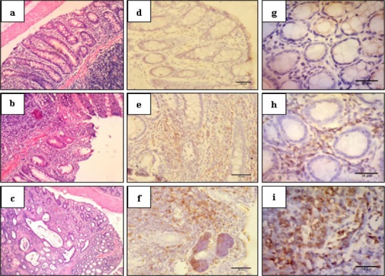
Figure 1.

Haematoxylin-Eosin and Immunohistochemical Staining of Colon Tissue in 400X Magnification. The histopathological of colon was considered as normal, colitis, dysplasia and adenocarcinoma (a-c). APC localisation was shown in stromal, stromal-epithelial border, epithelial and tumour cells (d-f). iNOS localisation was shown in stromal and epithelial cells (g-i). Normal colon (a) was not expressed APC (d) and iNOS (g). Colitis colon (b) expressed APC and iNOS in the cytoplasm of stromal. Dysplasia and adenocarcinoma colon (c) expressed APC in the cytoplasm of stromal, stromal-epithelial border and epithelial cells (f), and expressed iNOS in tumour cell, whereas iNOS expression was in the cytoplasm of tumour cell (g).
