Fig. 5.

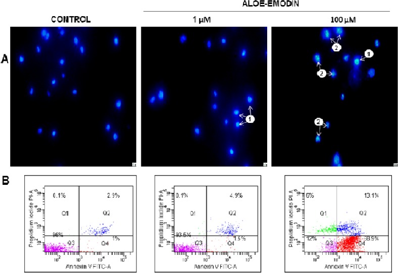
Apoptosis observed in HeLa cells using 4’,6-Diamidine-2-phenylindole staining (A). Control cells (not exposed to the test agent) showed normal morphology of the nucleus. After 48 hours of aloe-emodin action at the concentration of 1 µM, mainly cells with chromatin condensation were observed (1), whereas among cells exposed to aloe-emodin at the concentration of 100 μM numerous cells with both condensation (1) and nucleus fragmentation (2) were observed. Magnification × 400. Representative cytograms showing apoptosis analysis in a flow cytometer (B). Cells were treated for 48 hours with 1 μM and 100 μM concentration of aloe-emodin and apoptosis level was assessed by annexin V-FITC/PI staining. Control cells (not in apoptosis)-without Annexin V-FITC and PI staining. After the action of aloe-emodin, three populations of cells were demonstrated: living cells (Annexin V-FITC-/PI-), cells in the early (Annexin V-FITC +/PI-) and late stage of apoptosis (Annexin V-FITC+/PI+) and dead cells (Annexin V-FITC-/PI+). Data are representative of three parallel experiments.
