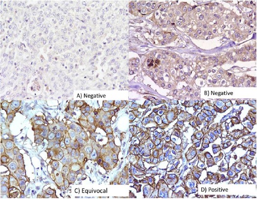
Figure 4.

Immunohistochemical Analysis of HER2 in Breast Carcinomas. The photographs show (a) negative control showing no detectable HER2 immunoreactivity in which HER2 antibody has been replaced with isotype specific IgG. (b) Equivocal and (c) Positive expression (A-D, original magnification ×200).
