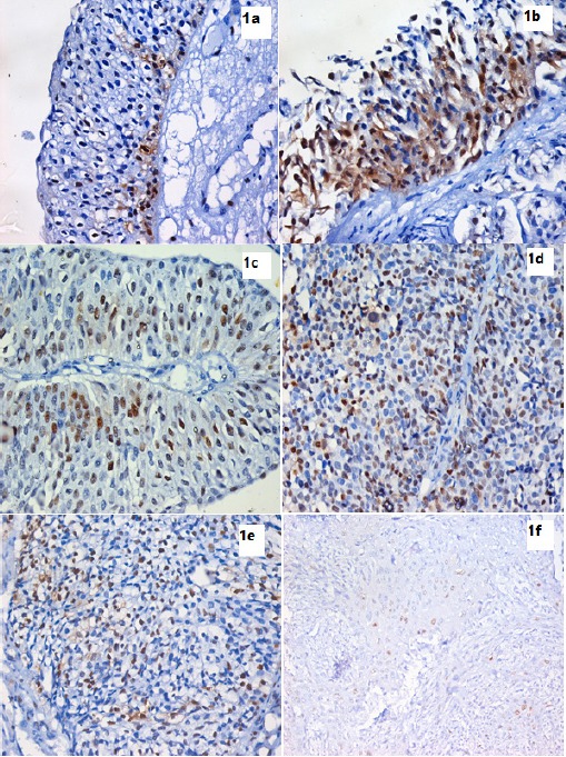
Figure 1.

IHC Using Anti-Cyclin D1 Monoclonal Antibody and DAB in Bladder Tissue Sections Expressed as Brown Mostly Nuclear Staining. 1a. Full-thickness urothelium in case of chronic cystitis showing positive nuclear and cytoplasmic expression of cyclin D1 in the basal layer. (IHC stain for cyclin D1, X400). 1b. Full-thickness urothelium in case of CIS showing positive nuclear and cytoplasmic expression of cyclin D1 in all layers. (IHC stain for cyclin D1, X400). 1c. Low grade superficial papillary UC showing moderate positive nuclear expression of cyclin D1. (IHC stain for cyclin D1, X200). 1d. Higher grade of non muscle invasive papillary UC showing increased positive nuclear expression of cyclin D1 compared to previous photo. (IHC stain for cyclin D1, X400). 1e. Non papillary muscle invasive UC showing lower positive nuclear expression of cyclin D1 compared to non invasive UC. (IHC stain for cyclin D1, X400). 1f. Section in a case of invasive SCC showing weak positive nuclear expression of cyclin D1. (IHC stain for cyclin D1, X400).
