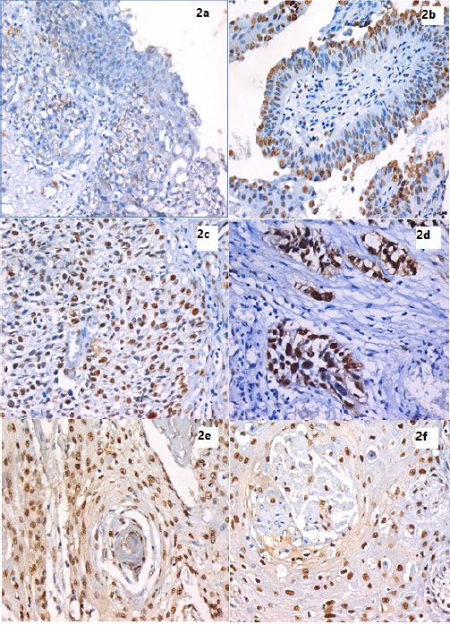
Figure 2.

IHC Using Anti-hnRNP-K Monoclonal Antibody and DAB in Bladder Tissue Sections Expressed as Brown Nuclear Staining. 2a. Full-thickness urothelium in case of chronic cystitis showing mild positive nuclear expression of hnRNP-K scattered through all layers. (IHC stain for hnRNP-K, X400). 2b. A case of Low grade superficial papillary UC showing mild positive nuclear expression of hnRNP-K mostly in the superficial layer. (IHC stain for hnRNP-K, X200). 2c. High grade papillary UC showing higher hnRNP-K expression than previous low grade superficial tumor . (IHC stain for hnRNP-K, X400). 2d. High grade non-papillary, muscle invasive UC showing marked hnRNP-K expression. (IHC stain for hnRNP-K, X400). 2e. A case of Bilharzial-associated SCC, showing high hnRNP-K expression. (IHC stain for hnRNP-K, X400). 2f. A case of non-bilharzial-associated SCC, showing high hnRNP-K expression. (IHC stain for hnRNP-K, X400).
