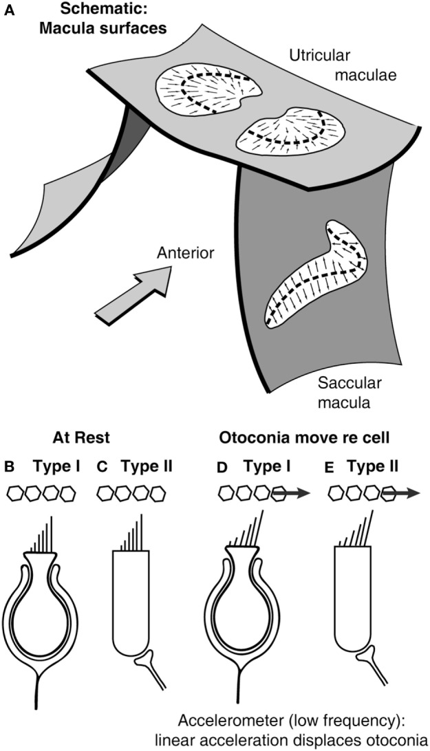
Figure 2.

(A) Schematic representation of the plates of otolithic receptors (the utricular and saccular maculae). The arrows show the preferred polarization of hair cell receptors across the maculae. The dashed lines are lines of polarity reversal (lpr). The striola refers to a band of receptors on either side of the lpr (27). Schematics of type I (B,D) and type II receptors (C,E) show how linear acceleration acts on otoliths and so deflects the hair bundles of individual receptors.
