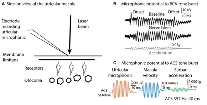
Figure 5.
Microphonic recording and laser Doppler measurement of macula movement, showing the relation between the vestibular microphonic and the velocity of macula movement during bone-conducted vibration (BCV) or air-conducted sound (ACS) stimulation. A microelectrode on the surface of the utricular macula (A) records a microphonic potential from the utricular receptors in response to BCV (B) or to ACS (C). There is no contribution from the cochlea since it has been completely ablated. (B) Vestibular microphonic responses to a BCV tone burst (40 ms, 400 Hz sinusoid) prior (top trace) and following (middle trace) lignocaine application to the vestibule to block the vestibular nerve. The microphonic remains after lignocaine injection showing it is a receptor field potential. Bottom trace: the BCV stimulus; the linear acceleration as recorded by a triaxial linear accelerometer on the ear bar. (C) Laser Doppler vibrometry. A laser beam is projected onto a reflective glass bead on the macula and the Doppler shift of the wavelength of the reflected beam shows the velocity of macula movement during BCV or ACS stimulation. Panel (C) shows the simultaneous measurement of vestibular microphonic and macula velocity. (B) Reprinted from Pastras et al. (57), © 2017, with permission from Elsevier. Panel (C) is from Pastras et al. (59).

