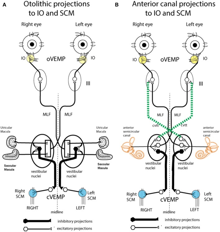
Figure 8.
Schematic representations of the major neural projections from vestibular receptors to the eye muscles and the neck muscles. (A) The otolithic projections to inferior oblique eye muscle (IO) and sternocleidomastoid muscle (SCM). (B) The analogous projections of the anterior semicircular canal neurons to the IO and SCM (72). Stimulation in animals with intact labyrinths causes the neural connections shown on the left panel to be activated. However, after a semicircular canal dehiscence (SCD), the anterior semicircular canals are also activated by sound and vibration, so the neural projections on the right come into play. The green dotted lines represent the projection from the anterior canal neurons in the vestibular nucleus to the contralateral third nerve nucleus via the crossed ventral-tegmental track. It appears that it is this combination of otolithic and canal afferent activation which in part results in the enhanced ocular vestibular-evoked myogenic potential (oVEMP) and cervical vestibular-evoked myogenic potential (cVEMP) responses after SCD. (A) Reprinted by permission from John Wiley and Sons, Curthoys et al. (80), © 2011. (B) Reprinted by permission from Springer Nature, Curthoys (81), © 2017.

