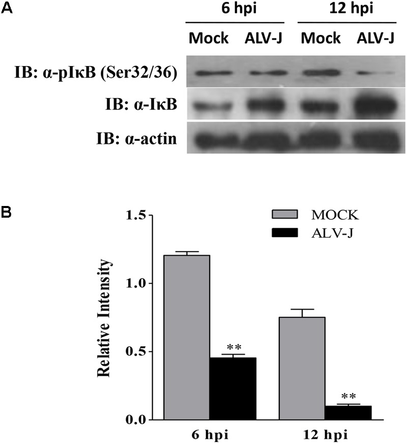
FIGURE 8.

ALV-J blocked the phosphorylation of IκBα on Ser32/32. (A) HD11 cells were infected with ALV-J or medium as a control. Cell lysates were prepared at 6 and 12 hpi, and examined by Western Blot using anti-pIκBα (Ser32/36), anti-IκBα and anti-actin antibodies. (B) Relative levels of IκBα phosphorylation in HD11 cells. The relative levels phosphorylated IκBα were calculated as follows: (density of bands of phosphorylated IκBα/band density of β-actin)/(density of bands of IκBα/band density of β-actin). Data are represented as means ± SD. ∗∗P < 0.01.
