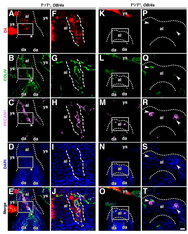
Figure 11. TC/TC mutant allantoises lack AX contribution and its derivative rod-like structure, which may organize the allantoic vasculature.

For all panels, the allantoic rod and PECAM1 midline vessel are encased by dashed lines, the allantois is outlined by dotted lines, and the proximal ACD is indicated with a white asterisk. (A-T) Frontal optical sections through the fetal-placental interface of a wildtype T+/T+ allantois (A-E) and a TC/TC mutant allantois (K-O) aided by higher magnification views of the white-boxed allantoic region in all of these panels (F-J, P-T), showing AX contribution to the allantoic rod (A-J) and failure of the TC/TC mutants to form a rod (K-T). White arrowheads (P-T), disorganized clusters of PECAM1- and COLIV-positive cells. Scale bar (T): 10 μm (F-J, P-T); 35 μm (A-E, K-O). al, allantois; da, dorsal aortae; ys, yolk sac.
