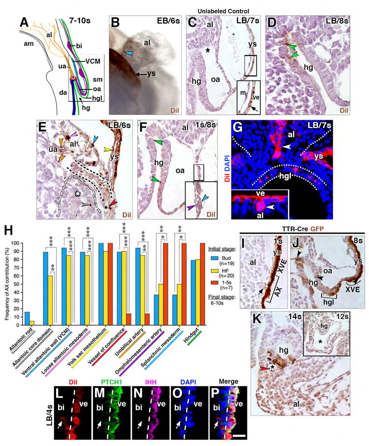
Figure 2. Contribution of AX to the fetal-placental interface.

(A) Structures encompassed within the fetal-placental interface, color coded to match results of fate mapping in panel H. Boxed region, structures that form a developmental continuum of those related to the nascent allantoic-yolk sac junction (see text). (B) Oblique ventral/side view, DiI-labeled whole mount specimen after culture and photobleaching shows contribution to the allantoic ventral wall (brown, blue arrowhead), visible after reflexion of overlying yolk sac (brown, background staining). (C-F) Sagittal histological sections through allantoic-yolk sac junction (E) and fetal-placental interface (C, D, F) showing a photobleached unlabeled cultured control (C) and cultured DiI-labeled specimens (D-F). Unlabeled photobleached control exhibits background staining limited to visceral endoderm (arrow in C inset, which is enlargement of main panel’s boxed region). Positive labeling (color-coded arrowheads): hindgut (D, F, green), umbilical artery (E, orange), loose allantoic mesoderm (E, pink), ventral allantoic wall (E, blue), yolk sac mesoderm (E, yellow), allantoic rod (E, white; outlined by black dashed line), allantoic core domain (E, grey, the region of which is indicated by white asterisk and outlined by white dashed line); vessel of confluence (E, red; black asterisk); splanchnic mesoderm (F, blue, inset of boxed region in larger panel); and omphalomesenteric artery (F, inset, purple). (G) Frontal optical section of the proximal-most region of the allantois of a DiI labeled specimen after culture with AX contribution to the allantoic rod-like structure (white arrowhead). The distal region, not shown, resembles the profile shown in panel E. Inset, rod-like structure (whitle arrowhead) in a transvers slice through reconstructed z-stack, at the level of white arrowhead in main panel. White dots outline allantois and hindgut lip. (H) Frequency of AX contribution to structures within the allantoic-yolk sac junction and fetal-placental interface; significance: Fisher’s exact test: *, P < 0.05; **, P < 0.01; ***, P < 0.001; the key along the X-axis is color-coded to match the color-coded arrowheads in panels C-G. (I-J) Sagittal (I, J) and transverse (K, and inset) histological sections through the fetal-placental interface of TTR-Cre-positive specimens (I-K) and a TTR-CRE-negative control specimen (inset, K) immunostained with anti-GFP. Brown staining is limited to Cre-positive specimens (I-J). Staining is continuous throughout extraembryonic visceral endoderm (XVE, I, J), including the AX (I), but it is mosaic in the hg (J, arrowheads; K) as well as in the hindgut lip (J, bracketed region). Arrow (I) indicates a mesodermal cell underlying the AX with slight brown staining. Brown staining is present in extraembryonic mesodermal tissues at 11-14s, including the VOC (asterisk, K; red arrowhead) and allantois (K). (L-P) Sagittal slice through reconstructed z-stack, DiI-labeled blood island-associated visceral endoderm co-localizes PTCH1 and IHH and makes negligible contributions to the mesoderm. Dashed line delineates yolk sac visceral endoderm. Scale bar (O): 10 μm (D, L-P); 15 μm (B, E, G); 20 μm (F, J); 35 μm (C); 43 μm (K, K inset). al, allantois; am, amnion; bi, blood island; da, dorsal aortae; hg, hindgut; hgl, hindgut lip; m, mesothelium; oa, omphalomesenteric artery; sm, splanchnic mesoderm; ua, umbilical artery; VCM, ventral cuboidal mesothelium; ve, visceral endoderm; ys, yolk sac.
