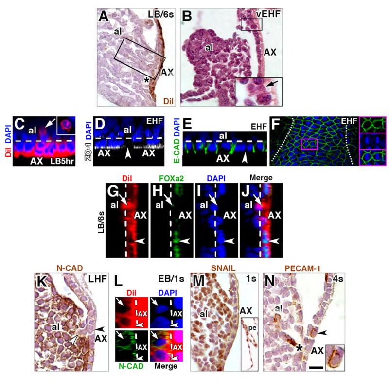
Figure 3. AX contributes to mesoderm via an epithelial-to-mesenchymal transition (EMT).

(A) Sagittal histological section, DiI-labeled cells streaming from the AX (boxed region) following culture and photobleaching. Black asterisk, vessel of confluence. (B) Sagittal histological section, allantoic bud, AX cell (arrow) extending basally into the allantois at the allantoic-yolk sac junction (enlarged in inset). The virtual space between allantoic bud and AX is a fixation and histological processing artifact. (C) Transverse slice, reconstructed z-stack of the AX, round DiI-covered/AX-derived cell (arrow, enlarged in inset from frontal optical section) that has been liberated from AX (delineated by the white dashed line) after 5 hours of culture. (D) Transverse slice, reconstructed z-stack, gap in axial continuity of ZO-1 (arrowhead) within AX (white dashed line). (E) Transverse slice, reconstructed z-stack, loss of junctional E-CADHERIN (arrowhead) between AX cells; white dashed line delineates the AX’s epithelium. (F) Frontal optical section, AX, E-CADHERIN-immunostained AX overlying the allantois (the extent of which is indicated by the white dots), highlighting mitotic profile in axial midline (magenta-outlined box), enlarged in insets on the right, showing individual channels (top, middle boxes) as well as merged image (bottom box). (G-J) Sagittal slice, reconstructed z-stack, of an exiting DiI-labeled AX-derived cell (arrow) which exhibits spotty FOXa2 staining; nuclei of remaining AX cells are robustly FOXa2-positive (arrowhead; AX, white dashed line). (K) Sagittal histological section, continuity between two N-CAD-negative cells, one of which remains within AX (black arrowhead) and the other has just departed (white arrowhead), entering the generally N-CAD-positive allantois. (L) Sagittal slice, reconstructed z-stack, exhibiting relatively little (bottom cell, arrowhead) or robust (top cell, arrow) N-CAD, while AX (dashed white line), is N-CAD-negative. (M) Sagittal histological section, SNAIL in allantois and parietal endoderm (inset), but not in AX or other visceral endoderm. (N) Sagittal histological section, rounded PECAM-1-positive AX cell (arrowhead, enlarged in inset). Black asterisk, VOC. Scale bar (N): 7 μm (C-E, G-J, L); 13 μm (K); 20 μm (B, N); 30 μm (F); 40 μm (A, M, M inset). al, allantois; pe, parietal endoderm.
