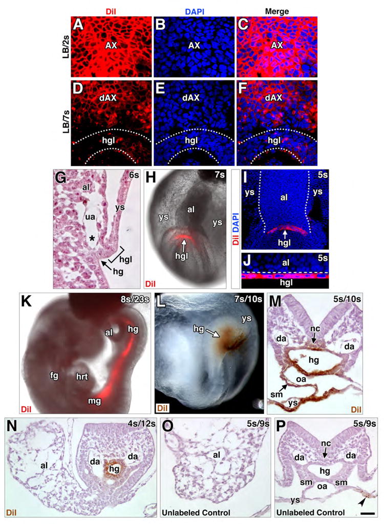
Figure 7. The hindgut lip, formed by the AX, is a mesendodermal tissue.

(A-F) Frontal (ventral) optical sections through AX (A-C) or dAX and hindgut lip (D-F). After short culture, DiI is undiluted in AX (A-C). After culture to later stages (D-F), dAX is largely uniformly labeled due to attenuation of its EMT, while the hindgut lip (hgl; outlined by dotted white lines), contiguous with the dAX, exhibits salt-and-pepper-like DiI, in accord with previous EMT activity and exhaustion of the label. (G) Sagittal histological section, fetal-placental interface, bracket defines the hindgut lip (hgl). (H) Oblique frontal view, whole mount specimen, fetal-placental interface, initial DiI label applied to hgl (red, white arrow). (I) Specificity of DiI to hgl in a frontal optical section of the fetal-placental interface (I) and a transverse slice through the hgl of the reconstructed z-stack (J). White dots (I) delineate the allantois; Dashed line (J) delineates allantoic cells from the hgl. (K) Right side view, whole mount specimen, DiI-labeled post-culture conceptus; fluorescent DiI visible within the midgut (mg) and hindgut (hg). (L) Oblique left-side view, DiI-labeled whole mount specimen after culture and photobleaching shows contribution to hg (brown, white arrow). (M-P) Transverse histological sections through fetal-placental interface (M, N, P) and allantois (O) showing cultured photobleached DiI-labeled (M, N) and unlabeled (O, P) specimens. Labeled specimens exhibit positive staining in the hg (M, N), notochord (nc, M), omphalomesenteric artery (oa, M) and splanchnic mesoderm (sm, M). Unlabeled photobleached control exhibits background staining limited to visceral endoderm (arrowhead in P); the allantois (O), hg (P), and other internal structures (P) exhibit no detectable staining. Scale bar (P): 10 μm (A-F); 20 μm (J, G); 40 μm (M, O); 50 μm (N, P); 75 μm (H, I, K); 100 μm (L); 145 μm (K). al, allantois; da, dorsal aortae; fg, foregut; hrt, heart; ua, umbilical artery; ys, yolk sac.
