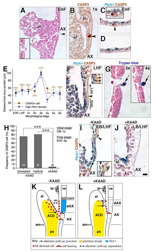
Figure 8. A single CASP3 cell, whose identity is regulated by Hedgehog signaling, separates non-EMT blood island visceral endoderm from the AX.

(A) Sagittal histological section, allantois, showing breach in the AX (arrowhead; region enlarged in inset). Breaches are not due to dissection, as embryos in which overlying parietal endoderm remained intact exhibited them (data not shown). (B) Sagittal CASP3-immunostained histological section, allantoic-yolk sac interface, displaying a single CASP3-positive cell (arrowhead) at the distal border of the dAX. (C, D) Transverse CASP3-immunostained histological sections, distal junction of the dAX in a Ptc:lacZ reporter conceptus. The unique CASP3-positive cell (C, arrowhead; enlarged in inset) is axially located within the midline and does not span into the consecutive section (D). (E) Graph displaying the co-location of the CASP3-positive cell and distal limit of the high Ptch1 dAX domain as the distance from the allantoic-yolk sac junction at increasing morphological stages. Sample sizes next to data points, along with the frequency at which the CASP3-positive cell was found for each stage. (F) Sagittal CASP3-immunostained histological section through the allantoic-yolk sac interface in a Ptc:lacZ reporter, CASP3-positive cell (arrowhead; enlarged in inset with arrowheads) is located between the visceral endoderm’s high Ptch1 domain and the yolk sac blood islands. Apically-localized punctate staining is visible in the visceral endoderm; this staining was commonly observed in extraembryonic visceral endoderm in this study. (G) Sagittal histological sections, yolk sac, showing several examples of trypan blue cells (arrows) being extruded from the epithelium at a similar distance from the allantoic-yolk sac junction as the CASP3 border cell (data not shown). (H) Frequency of the CASP3-positive cell in control (-KAAD) and KAAD-treated (+KAAD) specimens, sample sizes, base of each bar. Significance: Fisher’s exact test; ***, P < 0.001. (I, J) Sagittal CASP3-immunostained histological sections through the allantoic-yolk sac interface in cultured Ptc:lacZ reporter conceptuses; untreated (-KAAD, I), treated (+KAAD, J). While a CASP3-positive cell, just distal to the high Ptch1 domain, is found in the control (I, arrowhead; enlarged in inset), no signs of the high Ptch1 domain or the CASP3-positive cell are evident in the KAAD-treated specimen (J). (K, L) Schematic representation of the expanded AX domain in +KAAD specimens (L) relative to that in –KAAD (K). Scale bar (J): 9 μm (G); 15 μm (G inset, J); 20 μm (B, F, I); 25 μm (A, C, D). al, allantois; bi, blood island; hgl, hindgut lip.
