
Figure 9. Potency testing and/or fate mapping the ACD, ACD+AX, and AX via grafting.
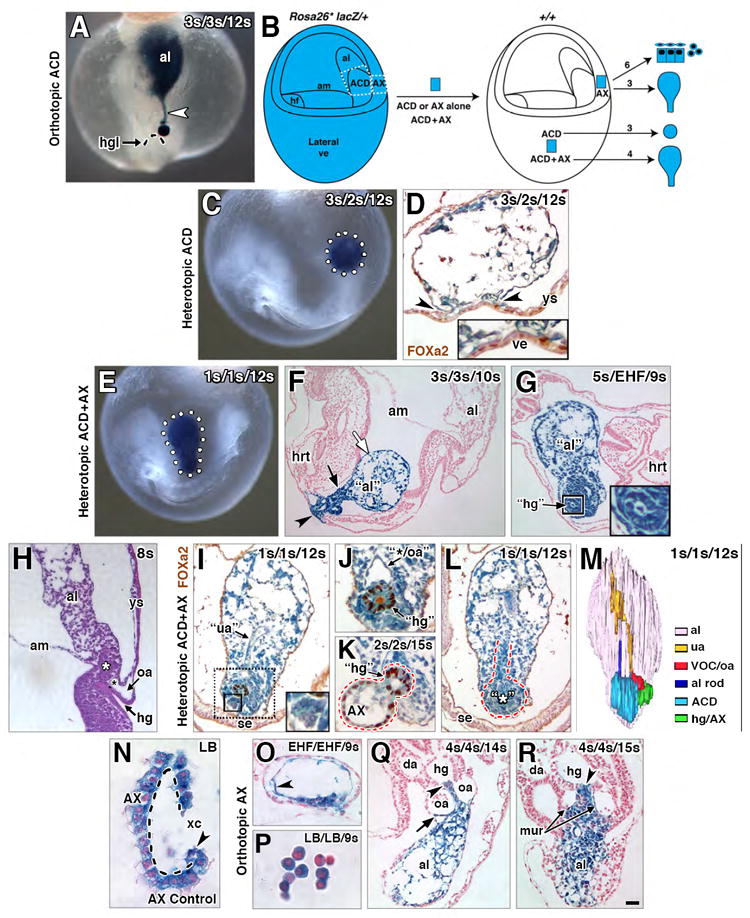
(A) Orthotopic (“same site”) ACD graft (blue) extending a midline file of cells (white arrowhead) from the hindgut lip (dashed arc) into the distal allantois. (B) Schematic diagram, grafting protocol. ACD, AX or ACD+AX were removed (white dashed regions) from a donor Rosa26*/+ conceptus and grafted into an F2 host conceptus. Lateral visceral endoderm (ve) was chosen as the heterotopic site as it was fairly tissue-free. Arrows, right of the host conceptus, outcome of each graft type (see text). Numbers above arrows, sample sizes. (C, D) Heterotopically-grafted donor ACD “sphere” (blue, dotted white circle) within host post-culture. (D) Transversely-oriented FOXa-2-immunostained histological section, donor ACD, and its contact with the lateral ve (arrowheads, higher magnification of this region, inset). The ACD is apolar, filled with disorganized small vessels, and lacks any indication of hindgut formation and/or the dense core domain. (E-G) Heterotopically grafted ACD+AX post-culture, whole mount preparations (E), showing the elongated shape (white dotted outline) of donor ACD+AX graft (blue). F and G, histological sections, integrated donor ACD+AX graft (F, blue, arrowhead), and a free-floating donor ACD+AX graft (G, blue color) within the host. Donor ACD+AX grafts resemble “stem-and-lollipop” structure of intact allantois and its connection to the hindgut; distal region loosely organized (white arrow, F); proximal region dense (black arrow, F) with a putative hindgut (boxed, G; enlarged in inset). (H) Sagittal histological section, fetal-placental interface, ex vivo conceptus showing the relationship between the distal and proximal regions in an intact conceptus. White asterisk, ACD; black asterisk, VOC. (I-L) FOXa-2-immunostained histological sections, heterotopic ACD+AX grafts (blue) post-culture. (I) Midline vessel, reminiscent of the umbilical artery (“ua”), runs through proximal half of the graft. At the proximal end (dotted black box), FOXa2-negative rosette formations (solid black box, enlarged in inset) are identified. (J) In the same specimen as (I), a single FOXa2-positive “hindgut” tube-like structure is adjacent to a large vessel, reminiscent of the VOC and/or omphalomesenteric artery (“*/oa”). (K) FOXa2 in the single “hindgut” structure and donor AX tissue (red dashed line). (L) Rod-like file of cells extends through the proximal midline of the graft from a dense core (red outline), reminiscent of the ACD (asterisk) and its rod-like extension. (M) 3-D model of free-floating graft whose colored structures are presented in panels I, J, and L. (N) Histological section through an uncultured control donor AX (dashed outline, basal side) clean of associated mesoderm. Arrowhead, apparent bilayer at this site is really the visceral endoderm curved on itself. (O-R) Histological sections through hosts and orthotopic AX grafts (blue) post-culture. (O, P) Bud and headfold-stage AX grafts, which failed to integrate into the hosts, formed squamous mesoderm (O, arrowhead) and a cluster of blood cells (P). (Q, R) Early somite-stage AX grafts, integrated into host, contributing to the allantois (Q, R), hindgut (Q, R, arrowhead), medial umbilical roots (mur, R), and the omphalomesenteric artery (oa) and overlying splanchnic mesoderm (Q, arrow). Scale bar (R): 10 μm (N, P); 20 μm (J, O); 30 μm (K); 40 μm (D, H, I, L, Q); 50 μm (A, R); 80 μm (C, G); 90 μm (E, F). al, allantois; am, amnion; da, dorsal aortae; hg, hindgut; hgl, hindgut lip; hrt, heart; mur, medial umbilical roots; oa, omphalomesenteric artery; se, surface ectoderm; ua, umbilical artery; VOC, vessel of confluence; ve, visceral endoderm; ys, yolk sac; xc, exocoelomic cavity.