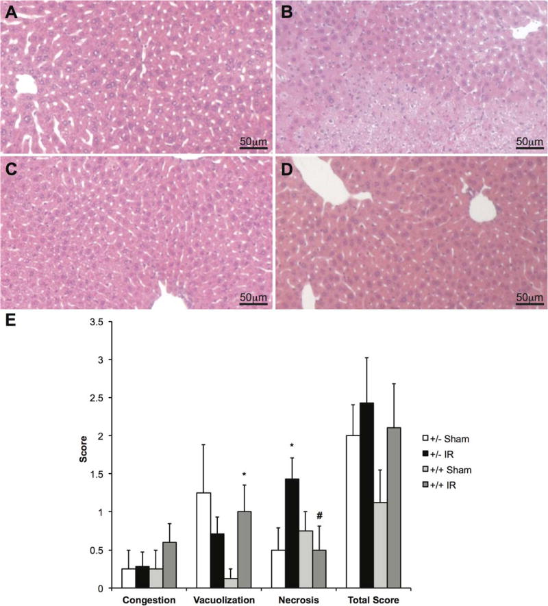
FIG. 3.

H & E staining of LysMcre/caNrf2 animals after partial hepatic ischemia. H & E staining of the LysMcre/caNrf2 animal livers was performed after either sham or partial hepatic ischemia surgery. Representative images for (A) LysMcre+/caNrf2− sham, (B) LysMcre+/caNrf2− IR, (C) LysMcre+/caNrf2+ sham, and (D) LysMcre+/caNrf2+ IR animals are shown. The sham group in both genotypes demonstrated normal liver histology. Although LysMcre+/caNrf2− animals after IR had large areas of necrosis, the LysMcre+/caNrf2+ animals after IR showed minimal necrosis. (E) The H & E slides were presented to 2 independent pathologists in a blinded manner, and Suzuki score was evaluated. On the basis of Suzuki scoring, the LysMcre+/caNrf2+ IR group demonstrated significantly decreased necrosis scores as compared to the LysMcre+/caNrf2− IR group. *P < 0.05 as compared to the sham group of the same genotype. # P < 0.05 as compared to +/− group of the same treatment.
