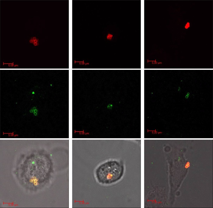
FIG 3.
Confocal microscopy images of infected HaCaT keratinocytes treated with CPP-JDlys. Keratinocytes were exposed to fluorescently labeled CPPTat-JDlys (green; middle row) and fluorescently labeled MRSA strain USA300 (red; top row) and monitored in real time by confocal microscopy. S. aureus bacteria were colocalized with CPPTat-JDlys (yellow) in the combined panels in the bottom row. Images of the three columns are representative of three independent experiments.

