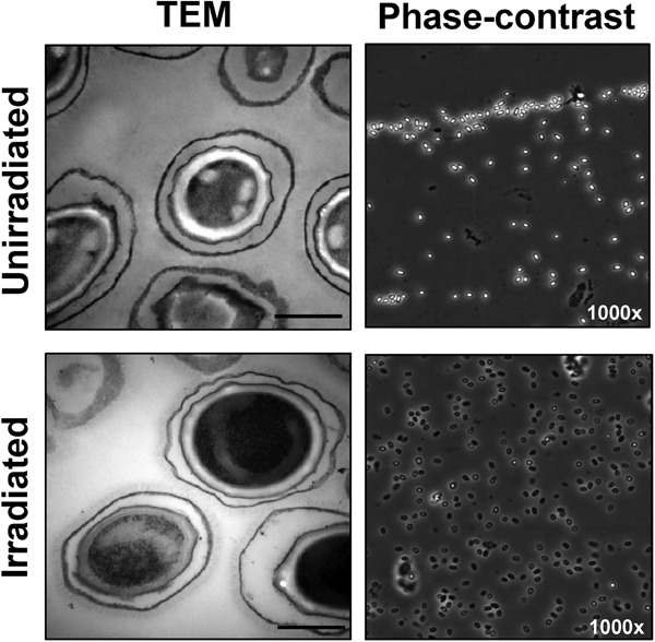
FIG 3.

Spore ultrastructure analysis. Irradiated and unirradiated B. anthracis Sterne spores were visualized by transmission electron microscopy and phase-contrast microscopy (50- and 40-kGy doses, respectively). Bars in electron microscopy represent 500 nm. Phase-contrast images were taken using an oil immersion objective at ×1,000 magnification.
