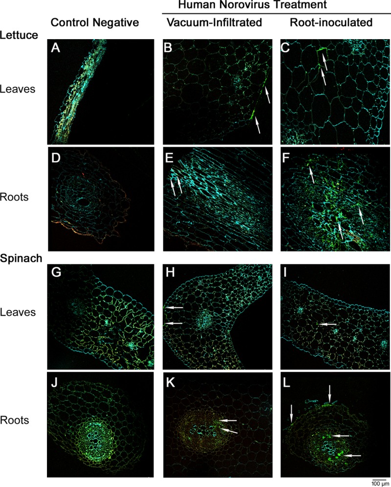
FIG 4.

(B, C, E, and F) Confocal microscopy images showing HuNoV inside leaves of lettuce plants that were vacuum infiltrated (B) or root inoculated with the virus (C) and their corresponding roots (E and F). (H, I, K, and L) HuNoV inside leaves of spinach plants that were vacuum infiltrated (H) or root inoculated with the virus (I) and their corresponding roots (K and L). (A, D, G, and J) Negative-control lettuce leaves (A) and roots (D) and spinach leaves (G) and roots (J). Examples of immunofluorescence associated with HuNoV detection are indicated by arrows.
