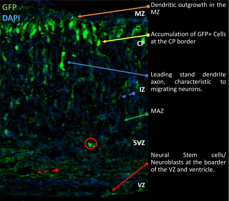
Figure 1.
Gross morphological schematic of sub-compartments in the developing rodent cortex. Representative image of developing cortex. Electroporation of eGFP was performed at E14.5 and brains collected at P0 as previously described (Srivastava et al., 2012a,b). The cortex is comprised of four morphologically distinct regions, the VZ, SVZ, IZ, and CP. Further to this there are the MAZ and MZ, located in the IZ and CP respectively. Located on the basal surface of the cortex proximal to the cerebral ventricles is the VZ responsible for generation of NSCs. Beyond the VZ, the SVZ contains proliferating and early differentiating neural progenitors. Between the SVZ and IZ, the MAZ is a point of accumulation of polarizing cells. After which the cells migrate through the IZ to the CP where terminal translocation takes place. This brief outline is the general schematic throughout development of the cortex. Cells migrate to the outmost layer and continually build on top of each other in a sedimentary manner. IZ, intermediate zone; MAZ, multipolar cell accumulation zone; CP, cortical plate; GFP, green fluorescence; NSCs, Neural Stem Cells; VZ, ventricular zone; SVZ, subventricular zone; MZ, marginal zone.

