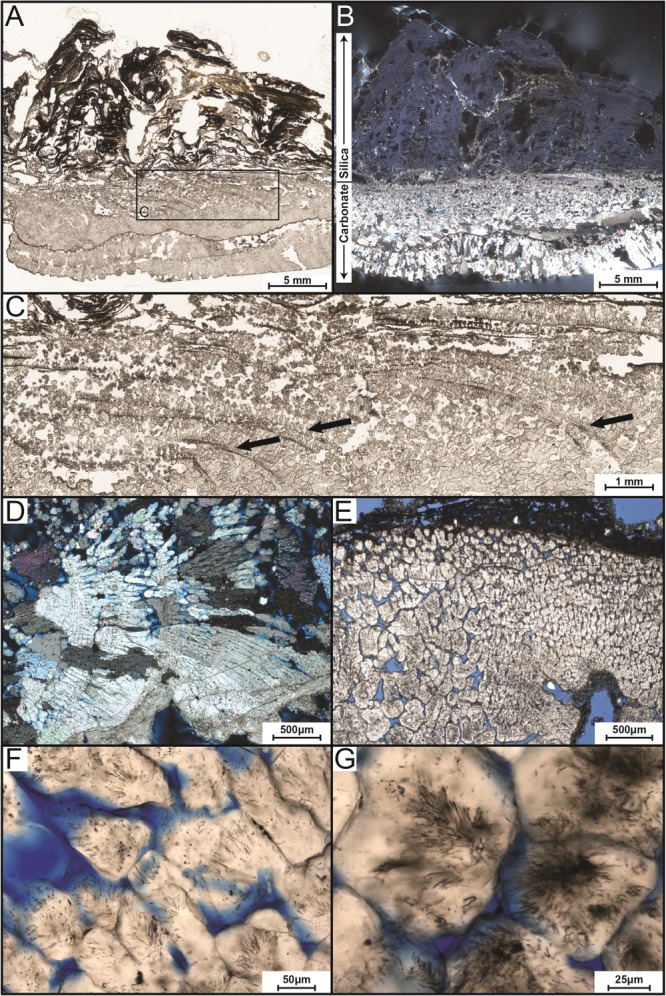
FIGURE 3.

Transmitted-light petrographic photomicrographs of LHC precipitates in thin section. (A) Plane-polarized transmitted light photomosaic of LHC precipitate (inset box enlarged in C). Light-colored area toward the bottom of the image is calcite and wavy-laminated, darker area toward the top is amorphous silica. (B) Cross-polarized transmitted light photomosaic revealing bladed calcite crystals in lower portion and amorphous silica in the upper portion (same field of view as A). Amorphous silica is opaque under cross-polarized light, so a minor amount of reflected light was cast across the thin section to enhance the visibility of the upper silica portion. (C) Inset image from (A). Arrows point to several curved, downward dipping surfaces that indicate progressive growth of the bladed calcite from left to right. (D) Crossed-polarized photomicrograph of bladed calcite from the lower portion of the precipitate indicating growth from left to right. (E) Plane-polarized transmitted light image of bladed calcite crystals with cloudy appearance caused by pervasive endolithic activity (blue areas represent pore space). (F,G) Plane-polarized transmitted light photomicrographs of endoliths within calcite crystals.
