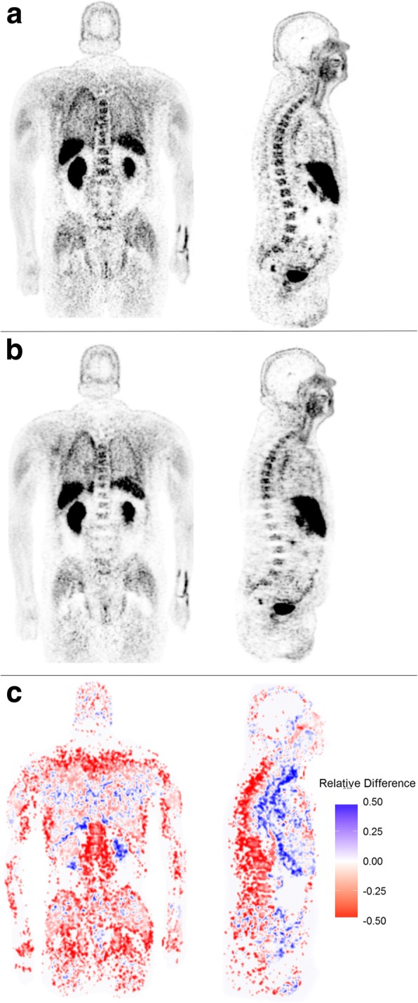
Fig. 2.

Coronal and sagittal TOF 18F-choline PET reconstruction (a). Coronal and sagittal non-TOF 18F-choline PET reconstruction (b). Relative percentage difference image (TOF - non-TOF)/TOF (c). Note the differences in the vertebra, the pelvic region, and the femora
