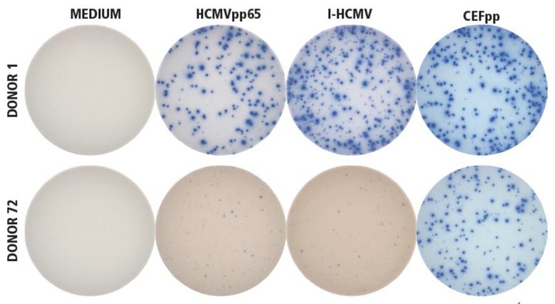
Figure 1.
Establishing the specificity of I-HCMV- and HCMVpp65-induced IFN-γ ImmunoSpot® formation. Donor 1 was seropositive for HCMV, Donor 72 was seronegative. PBMC from both donors were tested in an IFN-γ ImmunoSpot® assay in medium alone as the negative control (“MEDIUM”), or in the presence of the HCMVpp65 peptide pool (HCMVpp65) or inactivated HCMV virions (I-HCMV), as specified. As positive controls for eliciting memory T-cell responses, a pool of 32 peptides of HCMV, EBV, and influenza virus were used (CEFpp). The test was performed as specified in Materials and Methods, with three replicate wells for each condition. A representative image of one of the replicates is shown for each condition.

