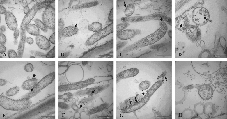
Fig. 4. TEM images of Las and phage particle morphology after 45 °C heat stress treatment.
a Control image of Las in periwinkle at 23 °C; phage particles are absent; b, c phage particles are visible 8 h after 45 °C temperature increase in Las-infected periwinkle; d, e phage particles are visible both inside and outside Las cells 3 days after 45 °C temperature increase; f, g phage particles released from Las cells 3 days after 45 °C temperature increase; h lysis of Las cells concurrent with release of phage particles 3 days after 45 °C temperature increase

