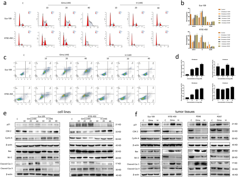
Fig. 5. Gimatecan induces S-phase arrest and apoptosis in ESCC.
a Eca-109 and KYSE-450 cells were treated with different concentration (10 nM, 20 nM, and 30 nM) of gimatecan and irinotecan for 4 h. Cell cycle progression was assessed using propidium iodide staining detected by fluorescence activated cell sorting. b Sums of percentages of each cycle were also calculated in Eca-109 and KYSE-450. Results are representative of three independent experiments. c Eca-109 and KYSE-450 cells were treated with gimatecan and irinotecan at the indicated dose for 72 h and stained with Annexin V-PE/7-AAD. d Sums of percentages of early apoptosis (Q3) and late apoptosis (Q2) were calculated as total apoptosis ratios. Results are representative of three independent experiments. e, f The expressions of proteins related to the cell cycle and apoptosis were assessed by Western blotting in vitro and in vivo. Eca-109 and KYSE-450 cell lines were exposed to 10 nM, 20 nM, and 40 nM concentration of gimatecan and irinotecan for 48 h, and harvested at 70–80% confluence. At the end of in vivo treatment, the mice were sacrificed and tumor tissues of Eca-109 and KYSE-450 cell line xenograft and PDX models were harvested. Cell cycle-related proteins, such as Cyclin A, CDK2, and p21, and Pro- and anti-apoptotic proteins including Bax, Bcl-2, cleaved-caspase 3, and cleaved-caspase 9 were assessed by Western blot. Data represent the mean ± SD of three replicate assays. *p < 0.05, **p < 0.01, ***p < 0.001

