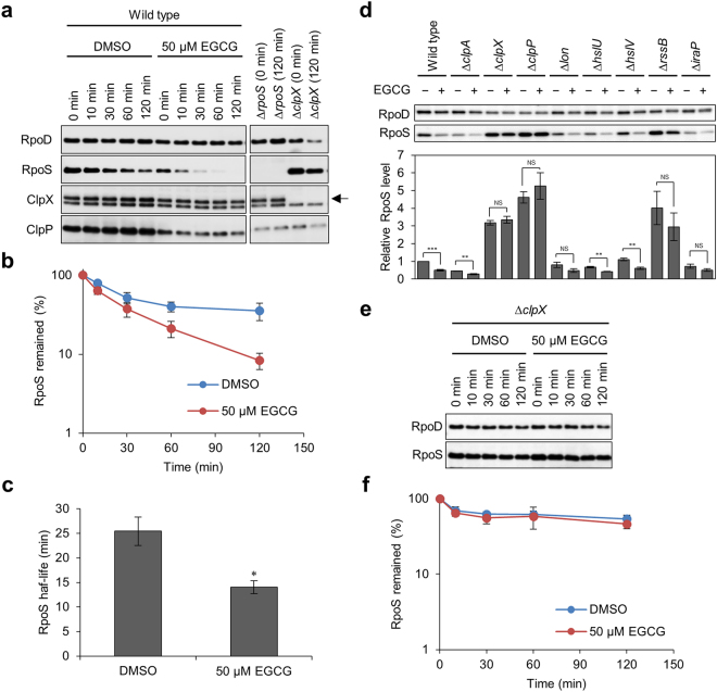
Figure 7.
Effects of EGCG on the stability of RpoS in vivo. (a) E. coli BW25113 wild type and its isogenic mutant cells (∆rpoS and ∆clpX) were grown to stationary phase (24 h) in YESCA medium supplemented with 50 μM EGCG. As a control, 1% DMSO was added to the medium. At various time points after the addition of Spectinomycin, cellular proteins were analysed by SDS-PAGE and immunoblotting with anti-RpoS, anti-RpoD (loading control), anti-ClpX, and anti-ClpP antibodies. In the panels of ClpX, the upper band indicated by an arrow corresponds to ClpX and the lower one is a non-specific protein. (b) Band intensities of RpoS in immunoblots in a were measured with the LAS-4000 Image Analyser. (c) Half-lives of RpoS in the presence of DMSO and EGCG were calculated from data in (b). The means and standard errors from at least triplicate determinations are represented. (d) Cellular RpoS levels in BW25113 wild type and mutant strains were analysed by immunoblotting as in a. These strains were harvested at 24 h without supplementation of Spectinomycin. Band intensities of RpoS in immunoblots were measured with the LAS-4000 image analyser. (e,f) Spectinomycin-chase experiments were also performed with ∆clpX. *P < 0.05; **P < 0.01; ***P < 0.001 (compared with DMSO control). Full-size scans of immunoblots are shown in Supplementary Figs S7–9.

