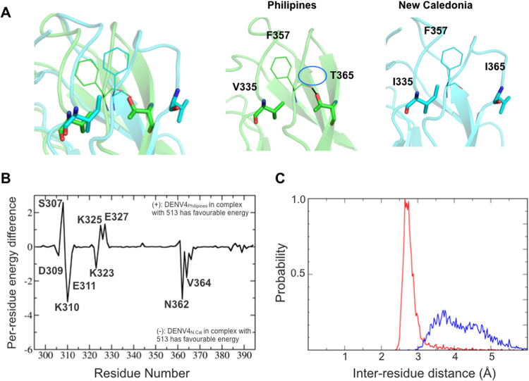
Figure 10.
Structural differences within DENV4 serotype between New Caledonia and Philippines isolates (A) Superimposed 3D structures of Philippines (green) and New-Caledonia (cyan) bound to 513 (not shown). The reorientation of F357 is shown in the left panel. The middle panel shows a representative snapshot from the trajectory, with the H-bond between the main-chain oxygen of F357 and OG1 of T365. The right panel shows the reorientation of F357 and I365. (B) Per-residue energy decomposition difference between DENV-DIII (DENV4 Philippines and New Caledonia) bound to 513; positive values indicate favorable binding energy of the residues from the Philippine strain while negative values indicate favorable binding energy for New Caledonia strain residues. (C) Plot of inter-residue distance between F357 and T365 (DENV4 Philippines strain in red) and F357-I365 (New Caledonia DENV4 strain, blue) over the equilibrated trajectories; the plot shows that the strong H-bond between F357:T365 maintains a more compact conformation, with an average bond length of 2.8 Å, while the corresponding conformation is loose in the DENV4 New Caledonia.

