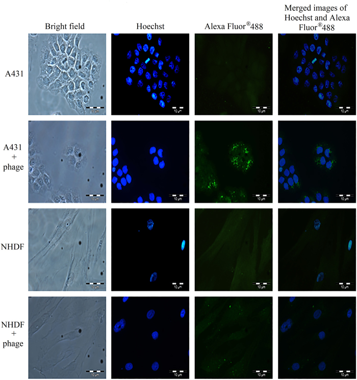
Figure 1.
Analysis of the internalisation of phage clone into A431 and NHDF cells with immunofluorescence microscopy. The cell lines used are labelled on the left. A431 and NHDF cells without phage served as negative controls. Purified phage (1 × 10¹² pfu) carrying the peptide NRPDSAQFWLHH was added to the cells. After incubation at 37 °C for 72 h, the phage was detected with the mouse anti-M13 monoclonal antibody, followed by the goat anti-mouse IgG conjugated to Alexa -Fluor® 488.

